
| Soluble Tumor Necrosis Factor Receptor 1 (sTNFR1) ELISA Kit | |
Alternative name:
|
|
| Receptor for TNFSF2/TNF-alpha and homotrimeric TNFSF1/lymphotoxin-alpha. The adapter molecule FADD recruits caspase-8 to the activated receptor. The resulting death-inducing signaling complex (DISC) performs caspase-8 proteolytic activation which initiates the subsequent cascade of caspases (aspartate-specific cysteine proteases) mediating apoptosis. Contributes to the induction of non-cytocidal TNF effects including anti-viral state and activation of the acid sphingomyelinase. The soluble form of human TNFR1 is produced from the membrane form by proteolytic processing. | |
Soluble TNF and IL-6 receptors: indicators of vascular health in women without cardiovascular disease |
|
| Cytokine receptor subunits are released from cells in a regulated manner and circulate in soluble forms at concentrations that are orders of magnitude greater than the concentrations of the cytokines themselves. The purpose of this study was to determine if the circulating concentrations of solublereceptor subunits for interleukin-1 beta (IL-1β), tumor necrosis factor alpha (TNFα) and interleukin-6 (IL-6) might serve as early indicators of vascular dysfunction independent of the traditional cardiovascular disease (CVD) risk factors in women. Healthy women, aged 20-50 years (n = 36), were assessed for circulating concentrations of the cytokines IL-1β, IL-6 and TNFα and the soluble cytokine receptor subunits interleukin-1 receptor type I (sIL-1RI), sIL-1RII, sIL-6Rα, glycoprotein 130 (s-gp130), soluble TNF receptor type 1 (sTNFR1), and sTNFR2, along with traditional CVD risk factors. Cytokine receptor subunit expression on mononuclear cells and the release of these subunits in vitro were also determined. Brachial artery flow-mediated dilation (FMD), carotid intima-media thickness (cIMT) and carotid-femoral pulse wave velocity (cfPWV) were assessed by ultrasonography and Doppler probes. Circulating sIL-6Rα correlated negatively with FMD (r = -0.56, p = 0.007) independent of age and other CVD risk factors. Circulating sTNFR1 correlated positively with cfPWV (r = 0.60, p = 0.002). TNFR1 receptor expression on monocytes correlated positively with cIMT (r = 0.51, p = 0.004). Plasma concentrations of IL-1β, IL-6 and TNFα were not significantly associated with FMD, cIMT or cfPWV. These data suggest that the receptors for IL-6 and TNFα, rather than the cytokines themselves, may be better indicators of early vascular changes that are associated with CVD. | |
| Cortez-Cooper M et al. Vasc Med. 2013 Oct;18(5):282-9 | |
Soluble TNF receptor 1-secreting ex vivo-derived dendritic cells reduce injury after stroke |
|
| Inflammation is a major factor in the progression of damage after stroke and in the clinic, current therapies treat the clot, not the resulting damage. We have developed a novel method of protein delivery that exploits the migration ability of leukocytes after ischemic stroke (transient middle cerebral artery occlusion; tMCAO). In our studies, ex vivo-derived dendritic cells (exDCs) migrate to the inflamed rat brain soon after tMCAO onset and the number of cells that remain in the brain after injection is significantly correlated with the amount of local inflammation at the injury site. In addition, exDCs transduced to overexpress soluble tumor necrosis factor (TNF) receptor1 (sTNFR1) produce functional cargo that is secreted and that blocks TNF-α bioavailability in vitro. When delivered at 6 hours post-tMCAO reperfusion, sTNFR1-exDC-treated rats show significantly smaller infarct size and decreased inflammation compared with animals treated with exDCs transduced with GFP lentivirus. These studies indicate that cell-mediated delivery of proteins may be a promising new approach to reduce brain damage after acute neurologic insult. | |
| Works MG, Koenig JB, Sapolsky RM.J Cereb Blood Flow Metab. 2013 Sep;33(9):1376-85. | |
|
Human Soluble TNFR1 ELISA Code No.: SK00111-02 Size: 96 T Price: $360.00 USD Standard Range:7.81-500 pg/ml Sensitivity:5 g/ml Sample Type: serum, EDTA plasma Sample requres: 25 ul, 100 ul per well of 10 fold diluted samples IntraCV: 6-8% InterCV: 8-10% Protocol: PDF |
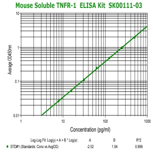 |
Mouse Soluble TNFR1 ELISA Code No.: SK00111-03 Size: 96 T Price: $360.00 USD Standard Range:7.81-500 pg/ml Sensitivity:5 g/ml Sample Type: serum, EDTA plasma Sample requres: 25 ul, 100 ul per well of 10 fold diluted samples IntraCV: 6-8% InterCV: 8-10% Protocol: PDF |
|
Human Soluble TNFR2 ELISA Code No.: SK00221-02 Size: 96 T Price: $360.00 USD Standard Range:7.8-500 pg/ml Dynamic Range: 7.8-500 pg/ml Sensitivity:7.8 g/ml Sample Type: serum, EDTA plasma Sample requres: 25 ul, 100 ul per well of 10 fold diluted samples IntraCV: 6-8% InterCV: 8-10% Protocol: PDF |


| Name | Code No. |
Size |
Price ($) |
| Human Soluble TNF-R1 ELISA Kit | 96 T |
360.00 |
|
| Human Soluble TNF-R2 ELISA Kit | 96 T |
360.00 |
|
| Mouse Soluble TNF-R1 ELISA Kit | 96 T |
360.00 |
|
| Mouse Soluble TNF-R2 ELISA Kit | SK00221-03 |
96 T |
360.00 |